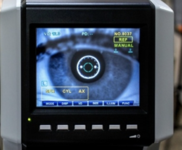
Dr George Hayek is involved in different areas of ophthalmology imaging and can give you several tests.
Retinal angiography is used to take background photographs after the injection of a dye (contrast agent) to visualize the retinal vessels and choroidal vessels. It is an examination that can detect vascular or inflammatory diseases of the eye.
Ocular biometrics is used to take different measurements of the eyeball in order to take the calculation for implanting a cataract or refractive surgery. The examinations are carried out by orthoptists and ophthalmologists and may require up to one hour of presence.
The examination of the visual field is used to explore the function of the optic nerve. It tests visual perception in different positions of space through stimulations with different light waxes. Examination of the visual field sometimes helps to detect other pathologies such as brain or neurological conditions.
Ocular echography allows diagnosis and follow-up of eye tumours. It uses ultrasound to explore the eyeball and orbit. We’re looking for potential retinal tears or retinal detachments.
The OCT per anterior segment is used to explore the anterior segment or the iridology-corneal angle of the eye. It allows an accurate analysis of the anatomy of structures: cornea, iris, crystalline, etc. The examination is performed in a sitting position without contact with the eye, it is performed without dilation of the pupil and it is recommended to avoid the make-up of the eye.
Corneal topography examination is very useful for detecting irregularities in curvature rays such as astigmatism which can be simple or complex, as a deformation of the cornea which can be progressive in the context of keratocin which is a contrindication to Lasik treatment to remove the correction by glasses.